MPS1
Live-cell imaging of Indian muntjac fibroblasts stably expressing H2B-GFP to visualize the chromosomes (green) and treated with 50 nM SiR-Tubulin to label spindle microtubules (magenta). Scale bar, 5 μm. Time, hr:min.
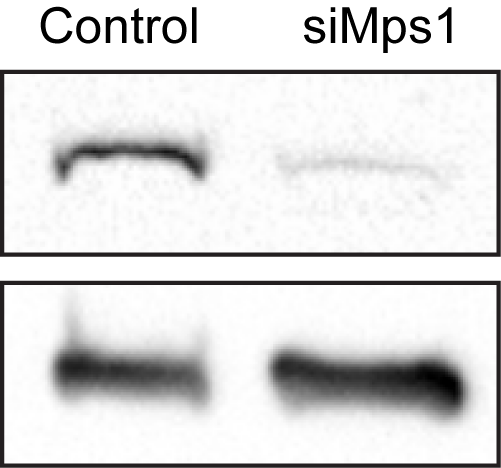
Protein lysates obtained after siRNA were immunoblotted with an antibody specific to each protein of interest. GAPDH was used as loading control.
Full WB here
Full WB here
Note 1: The cells were transfected with 50 nM of the target siRNA, 48h prior to live cell imaging/imunoblotting.
Note 2:
Note 2: